Heat-Treatment Assessment of a Greenish-Yellow Sapphire
APPLICATION CASE STUDY
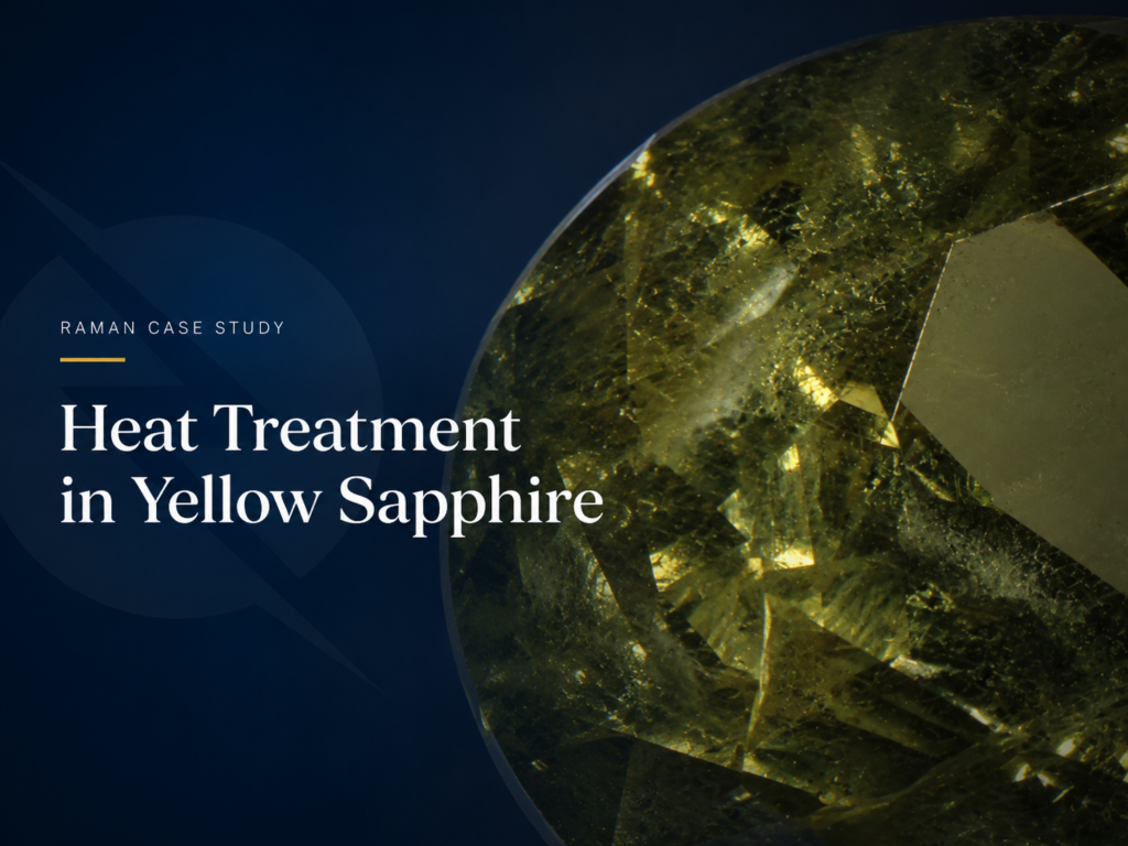
Heat-Treatment Assessment of a Greenish-Yellow Sapphire
A non-destructive multi-spectroscopic investigation of a 4.10 ct Madagascar sapphire using micro-Raman spectroscopy, photoluminescence and UV-Vis-NIR absorption, with complementary FTIR evidence.
STUDY AT A GLANCE
SPECIMEN
4.10 ct
Greenish-yellow oval sapphire
ORIGIN
Madagascar
9.64 × 7.88 × 6.03 mm
RAMAN
532 nm
30 × 5 s · 7 cm⁻¹ resolution
KEY RESULT
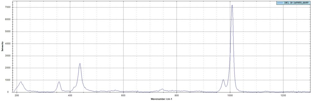
Zircon Identified
Bands near 215.5, 437.2 and 1008.1 cm⁻¹
MICROSCOPY & RAMAN
Targeted Identification of Internal Zircon Inclusions
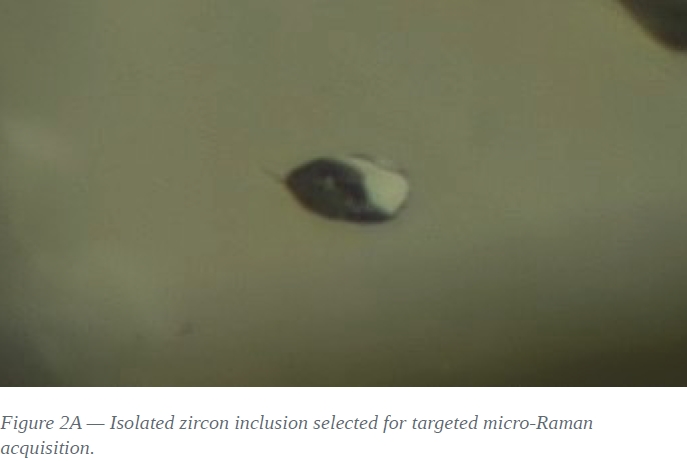
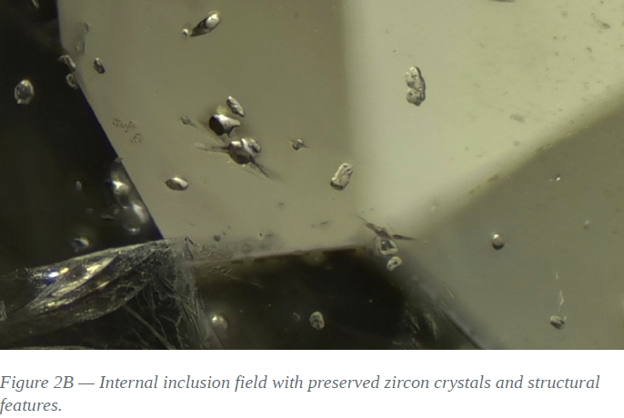
The Gurzil Raman Microscope combines optical microscopy for inclusion localisation with targeted micro-Raman acquisition. Zircon was identified spectroscopically and evaluated as one component of the heat-treatment assessment.
Raman confirms the mineral identity of the inclusion and contributes to the heat-treatment assessment as part of a convergent evidence framework. The measured FWHM should be interpreted as a supporting parameter rather than an isolated proof.
OPTICAL SPECTROSCOPY
Photoluminescence and UV–Vis–NIR Absorption
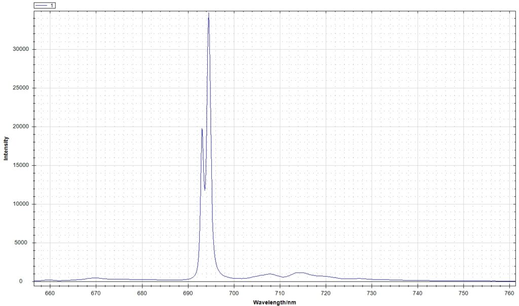
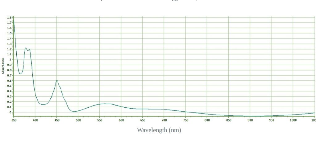
Photoluminescence resolves the Cr³⁺ R-line doublet, while UV–Vis–NIR absorption documents iron-related features and the broad visible absorption profile. Together, the two techniques provide complementary optical information on the corundum matrix and body colour.
The high-resolution PL spectrum resolves the Cr³⁺ R-line doublet, providing a clear optical signature of trace chromium in the corundum matrix.
UV–VIS–NIR ABSORPTION
Fe-Related Absorption Features
377 / 388 / 450 nm
350–1050 nm spectral range · UV–Vis–NIR Gemology Kit · broad shoulder near 560–580 nm
The absorption spectrum shows prominent Fe-related features and a broad visible shoulder, consistent with an iron-bearing sapphire and supporting interpretation of the greenish-yellow body colour.
COMPLEMENTARY FTIR EVIDENCE
Independent Supporting Evidence
Complementary FTIR measurements were performed on a third-party PerkinElmer Spectrum Two system. OH-related features in the 3000–3700 cm⁻¹ region were used as independent supporting evidence in the overall treatment assessment.
Complementary technique — not part of the Thunder Optics analytical platform.
DIAGNOSTIC ASSESSMENT
Convergent Multi-Technique Assessment
Each technique answers a different analytical question. The strength of the workflow lies in convergence: morphology, Raman mineral identification, optical spectroscopy and complementary FTIR evidence are considered together rather than as isolated proof points.
RAMAN
Zircon identified with Raman bands near 215.5, 437.2 and 1008.1 cm⁻¹.
OPTICAL SPECTROSCOPY
Cr³⁺ R-line emission and Fe-related UV–Vis–NIR absorption provide complementary information on the corundum matrix and body colour.
COMPLEMENTARY EVIDENCE
FTIR observations support the interpretation through OH-related features assigned to hydrous mineral phases
The combined observations are consistent with a natural sapphire showing no detectable evidence of heat treatment in the investigated specimen.
Explore the Thunder Optics Gemology Platform
This study combines Raman microscopy, photoluminescence and UV–Vis–NIR spectroscopy within a complementary analytical workflow for gemstone characterization.